News Story
UMD Biologist Awarded $1.5M to Develop Brain Mapping Techniques

Colenso Speer. Image courtesy of Colenso Speer.
The National Institutes of Health (NIH) awarded its Director’s New Innovator Award to University of Maryland Biology Assistant Professor Colenso Speer. The award, which is one of the organization’s most competitive grants, is part of NIH’s High-Risk, High-Reward Research Program. It is designed to support “exceptionally creative early career investigators who propose innovative, high-impact projects in the biomedical, behavioral or social sciences.”
Speer will receive $1.5 million for a five-year investigation into the development and plasticity of neural circuitry in the brain. He plans to create tools and methods for studying how neural circuits form and how connections between neurons change at the molecular level in response to sensory experiences.
Speer’s research could lead to a major step forward in mapping the physical layout of the circuits in the brain and understanding the complex chemical signals that pass between neurons.
“I was very pleased to receive this award and to know that our ideas are being welcomed and thought of as innovative,” Speer said. “This award acknowledges that success is not guaranteed, but that potential for a successful outcome is very high and that it would be transformative for the field.”
A key focus of Speer’s research is the molecular activity within synapses, the physical connections between neurons in the brain. Each of the roughly one hundred trillion synapses in the human brain is an extremely intricate and complex environment where multiple molecules process and convey signals across the connection. Those connections change over time, growing stronger or weaker through experience, a process that contributes to learning and memory. So far, scientists have not had the tools to investigate exactly how synapses function at the molecular level.
“We're sort of going down the rabbit hole in our research to try to understand how synapses are wiring up and properly connecting together as they’re being formed,” Speer said. “We’re asking, ‘What are the molecular mechanisms that regulate synapse formation and organization?’”
To conduct this research, Speer and his team will study how specific neurons and synapses develop to connect the eyes to the brain and enable visual perception in mice. Working with collaborators from UMD’s Department of Biology, Department of Chemistry and Biochemistry and Department of Cell Biology and Molecular Genetics, Speer will develop and apply a series of new technologies from three broad areas of research: transcriptomics, proteomics and super-resolution fluorescence microscopy. Transcriptomics, which analyzes RNA, will be used to determine how DNA in cells of the neuronal circuitry is transcribed to produce new proteins that perform the cells’ biological functions. Proteomics will be used to identify changes in the types and amounts of proteins at work in synapses during different stages of development and in response to different stimuli. Super-resolution fluorescence microscopy will enable researchers in Speer’s lab to image the physical arrangement of molecules within brain tissue and map the spatial organization of RNA and proteins contributing to the development of synaptic connections.
“The techniques that we plan to apply here are extremely sophisticated,” Speer explained. “We’re working with very small and delicate samples of RNA and protein that are either unstable, easily degraded or lost during the process, so each step is very technically challenging.”
In addition, the team will apply these methods to very tiny targets—single synapses from specific, individual neurons. With such a high level of difficulty, a lot of things could go wrong during sampling or analysis. But, if successful, Speer’s research could result in the first complete list of ingredients—at the level of RNA and protein—in a developing synapse. It could also provide the tools to create a full understanding of the spatial organization of the molecules that underly synapse function and the organization of circuits. This work would allow for a detailed molecular picture of how the brain is wired and how it changes in response to different sensory inputs.
“The reward is that we could have a comprehensive molecular map of synapses during development, aging and disease,” Speer said. “If we had that information, this would help us identify genetic candidates for potential treatments for neuropsychiatric disorders, neurodevelopmental disorders, aging and memory disorders, and many other cognitive functions that are directly impacted by changes in synapses.”
Speer came to the University of Maryland in 2017, after completing a postdoctoral fellowship at Harvard. He earned a Ph.D. in Neuroscience at UC Davis in 2010 and a B.S. in Molecular Biosciences/Biotechnology at Arizona State University in 2004.
###
Collaborators from the University of Maryland include Joshua Singer, a professor and chair of the Department of Biology, Peter Nemes, an associate professor of chemistry and biochemistry and Najib El-Sayed a professor of cell biology and molecular genetics who also holds a joint appointment in the University of Maryland Institute for Advanced Computer Studies.
Initial collaboration between Speer, Nemes and El-Sayed was supported by a 2019 UMD Brain and Behavior Initiative (BBI) Seed Grant.
Speer’s NIH New Innovator Award has been granted under the award number DP2MH125812.
Media Relations Contact: Kimbra Cutlip, 301-405-9463, kcutlip@umd.edu
This story originally appeared on the University of Maryland's College of Mathematical and Natural Sciences website.
Published October 6, 2020
Related Stories
Stories / June 14, 2022
Uncovering the mysteries of networking in the brain
Stories / October 14, 2021
Juntti Receives $1.9M Maximizing Investigators’ Research Award

Stories / July 7, 2021
MPower Seeks Proposals in Neuroscience and Aging, Medicine + AI

Stories / June 24, 2021
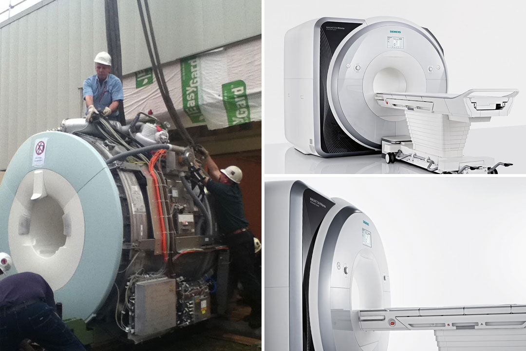
Piece of Mind: Upgrade of Campus MRI to Enhance Brain Imaging
Stories / May 17, 2021
Gumerov, Duraiswami Receive Funding from Facebook Reality Labs

Stories / March 29, 2021
Five UMCP-UMB Collaborations Awarded MPower BHHP Grants

Stories / March 22, 2021
El-Sayed to Direct New BBI Advanced Genomic Technologies Core

Stories / October 14, 2020
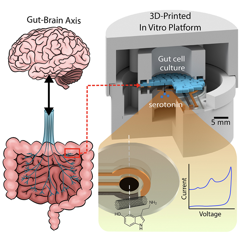
Symptoms all in your head—or in your gut? Maybe a little of...
Stories / January 31, 2020
Hu, Bernat Featured in Society for Women Engineers magazine

Stories / December 11, 2019
Brain and Behavior Initiative Hosts 3rd Annual Seed Grant...